Scanning Near-field Optical Microscopy
One of the possibilities of how to overcome the diffraction limit known from classical microscopy (the maximum resolution is
limited approximately by one half of the wavelength of the used light) is to use the scanning microscopy methods. With the help of SPM techniques
it is possible to bring the light source or detector of nanometric dimensions in a close proximity to the surface and break the diffraction limit.
Scanning Near-field Optical Microscopy (SNOM) is probably one of the most difficult SPM method considering not only the
experimental point of view (simultaneous force measurement, probe movement, excitation and optical signal acquisition) but also the data
interpretation. It was just the data interpretation that prevented a massive spread e.g. in biology where it was considered to be a promising
technique at the beginning.
From the experimental point of view we distinguish two basic regimes of SNOM:
|
-
In the aperture regime we use as a probe a optical fiber with very small aperture which is moved in a close proximity to the surface. The aperture is
typically 50-100 nm large which is also the highest possible lateral resolution. The fiber can be used either for sample illumination or for light
collection. The second part of the microscope (light collection or illumination) is usually made by a conventional optics but cases where
the fiber is used both for illumination and light collection also exist. For the detection of the force between the fiber and the surface a laser was
used in former devices similarly to the atomic force microscopy. However the presence of another laser decreases the signal to noise ratio;
in nearly all current devices we can find therefore a detection system based on the damping of vibration of a probe - piezoelectric excitation
element system where the excitation element is usually a tunning fork (e.g. from a watch crystal).
-
In the apertureless regime we use a metallized AFM probe which localy enhances the electric field in the vicinity of the probe tip. The probe makes
a scattering centre and we collect by a detector located in the far field the local light intensity. In order to minimize the influence of the light
scattering on the rest of the sample a synchronous detection connected to the z-axis AFM probe oscillation is used.
| 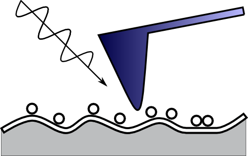 |
| Geometry of an apertureless measurement. For illustration, the light wavelength is
represented in proportion to the typicall surface roughness and nanoparticles size. |
Quantitative measurements
Despite the many years development in the SNOM field the quantitative analysis is still difficult. Certain approach can be based
on the use of a polarized light in order to set up a device similar to an ellipsometer. However it is necessary to say that the data analysis would
not also be easy. Within our research we try to perform numerical computations of electromagnetic field propagation in SNOM which could contribute
to better understanding of phenomena related to the image formation in this special microscopy technique.
|


